Stroke Vision Impact Calculator
Hemianopia
Loss of vision in the same side of both eyes. Often affects reading, walking, and driving.
Quadrantanopia
Loss of a quarter of the visual field. Can cause reading difficulties and spatial disorientation.
Visual Neglect
Failure to attend to one side despite intact vision. Often hidden by normal eye movements.
Cortical Blindness
Complete loss of conscious vision due to bilateral occipital lobe damage. May retain light perception.
When a stroke hits, most people think about speech or movement problems. But the eyes and brain’s visual system are just as vulnerable. Up to 70% of stroke survivors notice some change in what they see, and those changes can reshape everyday life faster than any other symptom.
Quick Summary
- Stroke can damage the visual pathway at many points, causing field loss, neglect, or total cortical blindness.
- Hemianopia (half‑field loss) is the most common visual deficit, affecting roughly one in three survivors.
- Visual neglect often hides behind normal eye movements, making it hard to spot without a specialist.
- Early eye‑brain testing and targeted rehabilitation improve functional recovery dramatically.
- Family members play a crucial role by adapting the home environment and encouraging regular visual exercises.
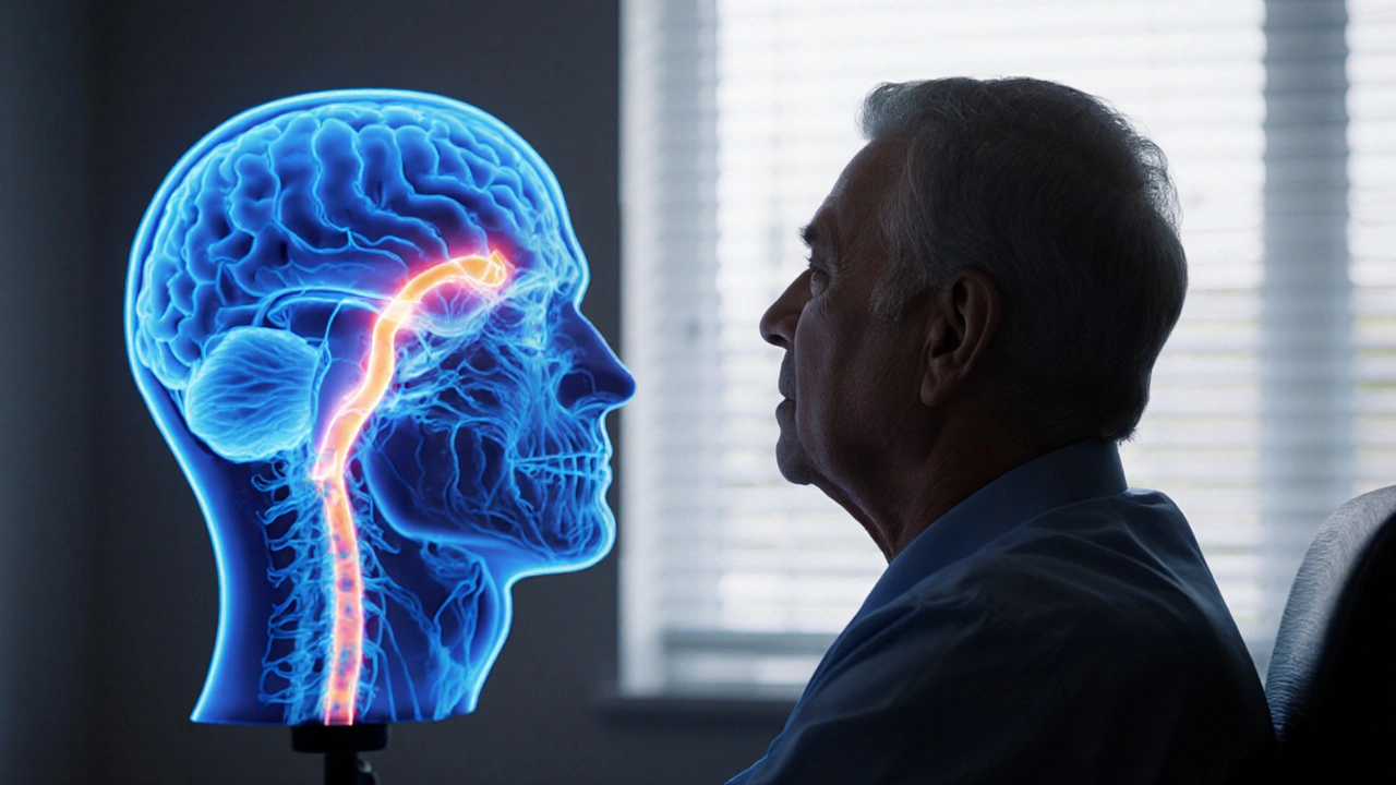
What Happens to the Visual System During a Stroke?
Stroke is a sudden interruption of blood flow to brain tissue, either from a clot (ischemic) or a bleed (hemorrhagic). When the visual cortex or its connecting pathways lose oxygen, the cells can’t fire correctly, leading to visual disturbances.
The visual system is a relay network that starts at the retina, travels through the optic nerves, crosses at the optic chiasm, and then splits into the optic radiations before reaching the primary visual cortex in the occipital lobe. Damage at any of these checkpoints produces a distinct pattern of loss.
The Most Common Visual Deficits After Stroke
Below is a quick snapshot of the four visual problems clinicians see most often.
| Deficit | Typical Brain Area Affected | Frequency Among Stroke Survivors | Primary Functional Impact | Rehab Focus |
|---|---|---|---|---|
| Hemianopia | Occipital lobe or optic radiations | ~30% | Missing half of visual field; bumping into objects | Scanning training, prisms, compensatory strategies |
| Quadrantanopia | Parietal or temporal optic radiations | ~10% | Loss of a quarter field; difficulty reading | Targeted eye‑movement exercises |
| Visual neglect | Right parietal lobe (left‑side neglect) | ~20% | Failure to attend to one side despite intact eyes | Awareness training, cueing, prism adaptation |
| Cortical blindness | Both occipital lobes | ~5% | Complete loss of conscious vision | Light perception training, adaptive tech |
Hemianopia: Half‑Field Blindness
Hemianopia is the loss of vision in the same side of both eyes-right or left. It occurs when the stroke hits the primary visual cortex (V1) or the optic radiations that carry signals from the retina.
Because the brain still receives input from the opposite side, patients often think they merely have a “blurred” area. The reality is they can’t see anything in that half, which makes driving, reading, and even walking precarious.
Recovery rates vary. A 2023 multicenter study found that 45% of hemianopic patients regained at least 20% of their lost field with intensive visual scanning therapy over six months.
Visual Neglect: The Brain’s Blind Spot
Visual neglect isn’t an eye problem; it’s a spatial attention issue. The eyes work, but the right parietal lobe can’t “tell” the brain to notice the left side of space.
Clinicians often spot neglect with the “line bisection test” - asking patients to mark the middle of a line. If they consistently err toward the right, neglect is likely.
Neglect dramatically slows recovery because patients may ignore a half‑filled plate or fail to see obstacles on one side, increasing fall risk.
Cortical Blindness: When the Whole Picture Fades
Cortical blindness occurs when both occipital lobes are damaged. Unlike optic nerve disease, the eyes and pupils react normally, making the condition tricky to diagnose.
Patients may retain light perception but lose shape and color discrimination. Some retain “blindsight” - they can respond to moving objects without conscious awareness, a phenomenon documented in neuro‑rehab literature.
While full visual recovery is rare, training to use residual light perception helps with navigation and confidence.
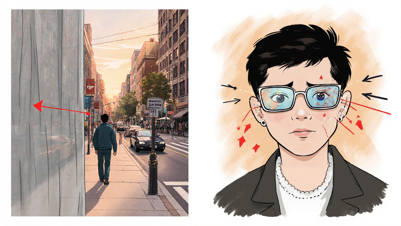
How Vision Changes Affect Everyday Life
Even a small field loss can turn simple tasks into hazards. Imagine reaching for a coffee mug that’s just out of sight, or crossing a street while missing a car on the blind side. These challenges often lead to anxiety, depression, and social withdrawal.
Family members may notice the survivor “turning their head” more often or “getting stuck” while dressing. Recognizing these cues early speeds up referral to specialists.
Getting the Right Diagnosis
The first step is a thorough eye‑brain assessment performed by a neurologist. The exam includes:
- Visual field testing (automated perimetry or confrontation test).
- Eye‑movement tracking to spot neglect or saccadic deficits.
- Brain imaging (MRI or CT) to pinpoint lesion location.
After the neurologist, an ophthalmologist evaluates the eye structures to rule out retinal or optic nerve disease that could mimic stroke‑related loss.
Early detection matters. The sooner therapy begins, the better the brain can reorganize and compensate - a principle known as neuroplasticity.

Rehabilitation Strategies That Work
Visual rehabilitation is a team effort. Here are the most evidence‑based approaches:
- Scanning training: Patients practice moving their eyes deliberately toward the blind side while performing daily tasks. Studies show a 30% improvement in reading speed after eight weeks.
- Prism glasses: Fresnel prisms shift the image from the blind field into the seeing field, allowing quicker hazard detection while walking.
- Computerized visual field restitution therapy (VRT): Repetitive light‑stimulus tasks stimulate the damaged cortex. Meta‑analysis (2022) reports modest gains for hemianopia.
- Neglect cueing: Using tactile or auditory cues on the neglected side can retrain attention networks.
- Adaptive technology: Screen‑magnifiers, high‑contrast keyboards, and voice‑controlled assistants reduce reliance on visual input.
Consistency is key - exercises performed 20‑30 minutes daily yield the best outcomes.
Managing Expectations and Emotional Health
Recovery isn’t always linear. Some patients plateau, while others regain function years later. Setting realistic goals helps keep motivation up.
Psychological support is essential. A 2021 trial showed that combining visual rehab with counseling cut depression scores by 40% compared to rehab alone.
Caregivers should:
- Remove tripping hazards and keep high‑traffic areas clutter‑free.
- Label drawers and cabinets with tactile markers.
- Encourage regular eye‑brain exercises, even when progress feels slow.
Key Takeaways for Patients and Families
Understanding the link between stroke and vision empowers everyone to act fast. The stroke vision impact is real, but with proper testing, targeted therapy, and a supportive environment, most survivors regain functional independence.
Frequently Asked Questions
Can vision improve after a stroke?
Yes. About 40-50% of people with hemianopia experience measurable field recovery with intensive visual scanning and prism therapy. Even those with cortical blindness can learn to use residual light perception for daily navigation.
How is visual neglect diagnosed?
Neglect is identified through behavioral tests such as line bisection, star cancellation, and the “copy” task. The clinician also observes whether the patient spontaneously attends to the neglected side during activities.
Do glasses help with post‑stroke vision loss?
Standard prescription glasses do not fix field loss, but prism glasses can redirect images from the blind side into the seeing side, improving safety while walking or driving.
Is there a risk of falling because of visual deficits?
Yes. Studies show that stroke survivors with untreated hemianopia or neglect have a 2‑3× higher fall rate. Early visual rehab and home modifications dramatically cut that risk.
How long does visual rehabilitation usually last?
Therapy is typically prescribed for 8-12 weeks, but many clinicians recommend ongoing home‑based exercises indefinitely to maintain gains.
Releted Post

Caspian Fothergill
Hello, my name is Caspian Fothergill. I am a pharmaceutical expert with years of experience in the industry. My passion for understanding the intricacies of medication and their effects on various diseases has led me to write extensively on the subject. I strive to help people better understand their medications and how they work to improve overall health. Sharing my knowledge and expertise through writing allows me to make a positive impact on the lives of others.
Hey everyone! 🌟 If you or a loved one are navigating vision changes after a stroke, remember you’re not alone. Tiny daily eye‑movement drills can add up to big improvements over weeks. Keep the environment well‑lit and don’t shy away from using prism glasses if they help. You’ve got this, and the community’s got your back!
Picture the brain as a bustling city, and a stroke as an unexpected blackout in the visual district. 💥 The aftermath isn’t just blurry text; it’s a full‑blown re‑routing of perception. Aggressive therapy, like scanning training, forces those neural detours to become highways. Embrace the colorful journey of relearning, because the mind loves a challenge! 🚀
Imagine being told that your brain has decided to turn half the world into a silent movie 🎬. The drama of suddenly missing a side of your vision is enough to make anyone feel like an actor on an invisible stage. Yet, the irony is that the eyes themselves are fine; it’s the brain pulling the curtain. Let’s not forget that rehab isn’t just medical-it’s theatrical, demanding applause for every tiny triumph. 💃
Oh come on, this whole "dramatic" thing is just a excuse for laziness. If you cant see half the screen you probbly didnt practice enough. Real american stoic would just push through and stop whinin about it. Stop looking for excuses and start training.
Post stroke vision loss is common and should be addressed early it helps with recovery and reduces risk of falls
Absolutely, early assessment is key. A simple visual field test can reveal a lot, and from there therapists can tailor scanning exercises that fit daily routines. It’s also helpful to involve family members in the training so they can provide cues and encouragement during regular activities.
Just a quick heads‑up: those little eye‑movement drills you hear about aren’t just filler-they actually train the brain to map missing spaces. 📚 I’ve seen friends swing from fearing stairs to confidently navigating their kitchen after consistent practice. Keep it chill, stay consistent, and celebrate the tiny wins! 😊
Yo dude that drill is sooo boring but hey if it works i guess.. .
For anyone starting rehab, focus on short, frequent sessions-about 20 minutes a day. Use high‑contrast cards to train scanning, and consider prism glasses if you have hemianopia. Consistency beats marathon sessions, and tracking progress with a simple chart can keep motivation high.
Your advice lacks proper structure and punctuation its riddled with slang and missing commas its not academically sound this is exactly why many patients get mis‑informed proper rehab requires evidence‑based protocols and precise language this post would benefit from citations and clearer formatting stop being vague.
It is an unfortunate truth that modern medicine often overlooks the profound psychological impact of visual deficits after a cerebrovascular accident. The loss of half a visual field or the subtle neglect of one side of space is not merely a functional inconvenience; it is a moral failing of our health systems that we allow patients to suffer in silence. When a survivor wakes each morning and cannot locate the coffee mug on the left side of the table, it is a stark reminder of the fragility of our neural circuitry. Ethical caregiving demands that we allocate resources not just for motor rehabilitation, but for comprehensive visual therapy that addresses both the physiological and existential crises. The brain, in its remarkable plasticity, can relearn to attend to the neglected periphery, yet it requires disciplined, evidence‑based interventions that are often underfunded. Moreover, the social stigma attached to visual neglect frequently leads to isolation, reinforcing depressive spirals that can undo even the most intensive physio work. Families must be educated to recognize these subtle cues, and clinicians should adopt a holistic approach that includes occupational therapists, neuro‑ophthalmologists, and mental health professionals. In my experience, the integration of high‑contrast environmental modifications and structured scanning exercises yields measurable improvements in daily functioning, but these outcomes are contingent upon persistent, patient‑centered care. The narrative of recovery cannot be reduced to a binary of “improved” or “not improved”; it must be a nuanced story that acknowledges setbacks and celebrates incremental gains. Therefore, institutions should mandate routine visual field assessments as part of post‑stroke protocols, ensuring early detection and timely referral. Only through such systematic vigilance can we hope to mitigate the profound loss of autonomy that visual deficits impose. The ethical imperative is clear: to treat the whole person, not just the limbs. This commitment will, in turn, reduce the societal burden of disability and improve quality of life for countless survivors.
Wow, a dissertation on vision loss, impressive 🙄. Guess we’ll all start quoting that at dinner parties now 😂.
That's just nonsense.
Clearly you missed the point. Visual deficits are well‑documented and the rehab protocols I use daily are proven to work. Stop spreading misinformation.